In a pioneering study utilizing magnetic resonance imaging (MRI), researchers have unveiled preliminary evidence indicating a potential connection between cognitive impairments and reduced pulmonary gas exchange in individuals experiencing Long COVID symptoms. This study, set to be presented at the Radiological Society of North America (RSNA) conference, sheds light on ongoing symptoms of breathlessness and fatigue in patients long after their initial COVID-19 infections.
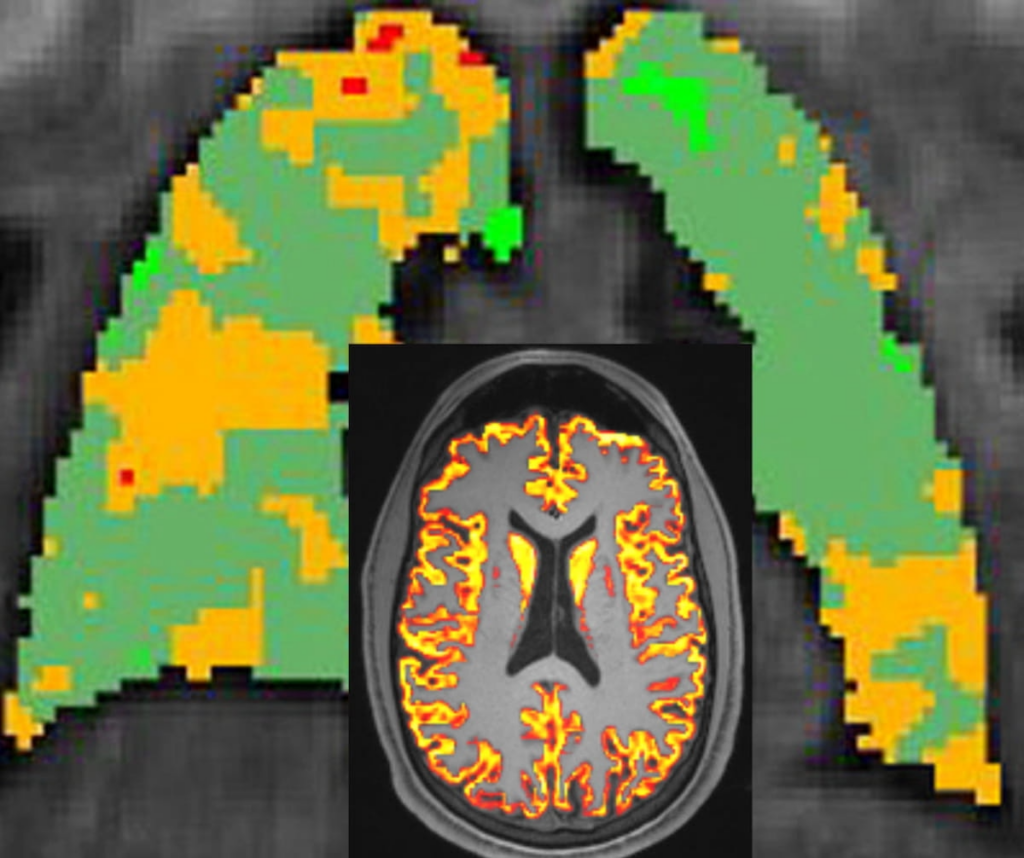
The research team delved into data from hyperpolarized 129Xe pulmonary MRI as well as structural and functional brain MRI for a group of 11 participants. These individuals had been grappling with enduring respiratory symptoms and fatigue approximately 31 months following their acute COVID-19 infections. The findings suggest a striking correlation: those with compromised pulmonary function tended to exhibit decreased white matter in the cerebral and parietal lobes, alongside diminished gray matter in the frontal lobes of the brain.
The study’s notable feature was the employment of hyperpolarized 129Xe pulmonary MRI, a tool deemed particularly valuable for this particular patient population. According to Keegan Staab, B.S., the lead author and a graduate research assistant in the Department of Radiology at the University of Iowa, this advanced imaging technique offers significant insights into ventilation and gas exchange processes. Staab emphasized that the current literature suggests that 129Xe may offer greater sensitivity to pulmonary injuries than standard respiratory tests, making it especially advantageous in studying Long COVID patients who often present with normal results in conventional breathing assessments.
The researchers also discerned an intriguing association between reduced pulmonary gas exchange and elevated cerebral blood flow. However, they urge caution, noting the necessity for more extensive studies to thoroughly explore this relationship. Staab theorized that this correlation might represent a compensatory response, where reduced lung function triggers an increase in cardiac output and brain perfusion. Alternatively, the disease mechanisms impeding pulmonary function might also contribute to increased brain perfusion through vascular damage affecting both the lungs and the brain.
This groundbreaking research highlights the complex interplay between respiratory and cognitive symptoms in Long COVID patients. As our understanding of this condition grows, studies like this one will be invaluable in guiding therapeutic strategies and improving the quality of life for those affected by persistent post-COVID-19 symptoms.
Reference: Stabb K, McIntosh M, Percy J. Xenon-129 MRI pulmonary gas exchange in long Covid is associated with cognitive function and brain MRI. Poster to be presented at the Radiological Society of North America (RSNA) 2024 110th Scientific Assembly and Annual Meeting Dec. 1-5, 2024.


