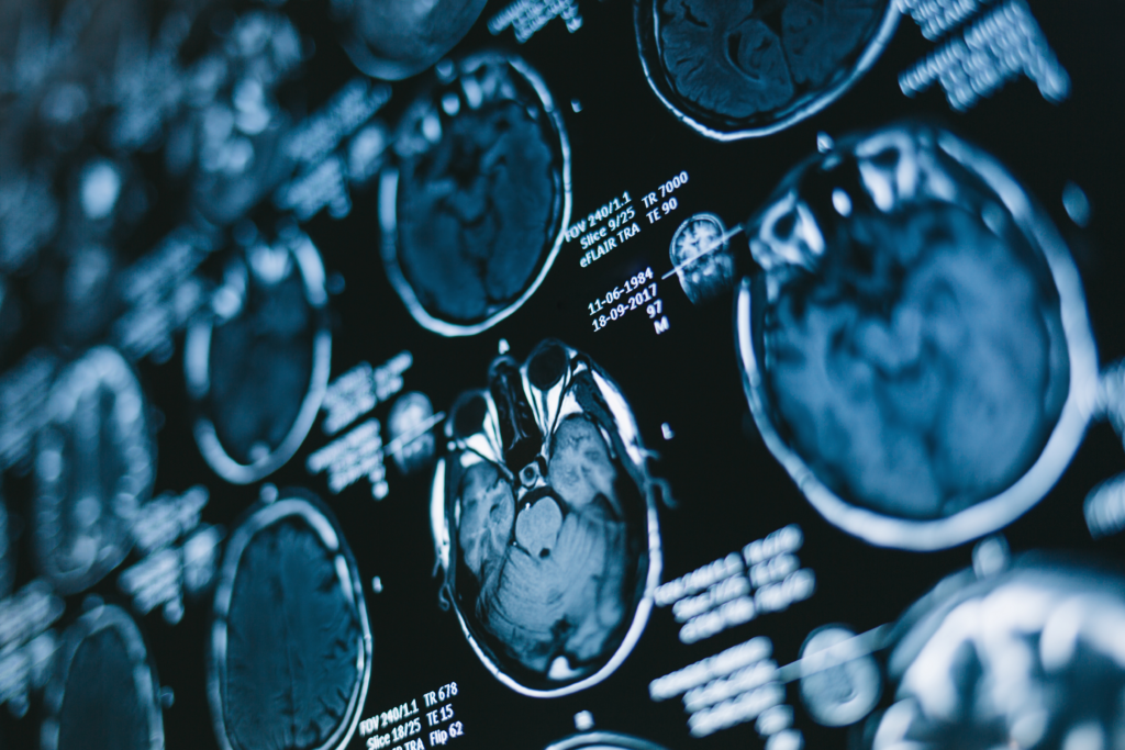
Recent research has indicated that for pre-operative evaluations related to grading and molecular characterization of diffuse gliomas, gadolinium-free magnetic resonance imaging (MRI) may be just as effective as gadolinium-based contrast agent (GBCA)-enhanced MRI. This finding comes from a retrospective study published in the journal European Radiology, where researchers employed a diagnosis prediction decision tree (DPDT) based on gadolinium-free MRI to analyze 303 patients with grade 2-4 adult-type diffuse gliomas.
The studied cohort included a mix of 54 patients with low-grade gliomas and 249 who had high-grade gliomas. Among these, 82 were identified as having IDH-mutant glioma, while 221 were classified as wildtype. Additionally, 34 patients exhibited a 1p/19q-codeleted glioma, with the remaining 269 having intact 1p/19q status.
The findings revealed that the accuracy of predicting glioma grades was greater than 85% using gadolinium-free MRI, closely matching the more than 87% accuracy achieved with GBCA-enhanced MRI. When it came to molecular status predictions, the results were also comparable, with gadolinium-free MRI showing around 75% accuracy compared to 77% accuracy for the GBCA-enhanced approach.
Dr. Aynur Azizova, the lead author of the study from the Radiology and Nuclear Medicine Department at Vrije Universiteit Amsterdam, expressed optimism regarding the effectiveness of gadolinium-free MRI. She noted that “our study suggests that the proposed DPDT using GBCA-free MRI could be as reliable as standard GBCA-enhanced MRI in preoperative diagnostic glioma assessment.”
Interestingly, while there was substantial overall agreement regarding molecular status predictions with both imaging methods (77% for GBCA-enhanced MRI to 75% for gadolinium-free), there was a notable difference in inter-rater agreement when evaluating tumor grades. The GBCA-enhanced MRI showed 68% agreement, which is 12% higher than the 56% agreement recorded for gadolinium-free MRI interpretations.
The researchers indicated that the DPDT model utilized 11 distinct imaging features drawn from the gadolinium-free scans. Specific characteristics, such as the T2-FLAIR mismatch sign and calcifications, were frequently observed in astrocytoma and oligodendroglioma cases. However, the study also highlighted discrepancies in agreement for assessments regarding midline shift and hemorrhage, suggesting potential inconsistencies that may stem from differing interpretations rather than the MRI techniques themselves.
For example, the variations in hemorrhage assessment may relate to the inconsistent availability of susceptibility-weighted imaging (SWI) and differences in pre-contrast T1 hyperintensity, rather than the absence of GBCA-enhanced images. Similarly, measurement disagreement for midline shift could likely arise from the challenge in accurately gauging minor shifts around a 5mm threshold, which leads to discrepancies among evaluators.
Significant Insights from the Study
-
Comparable Glioma Grading Accuracy: The study demonstrated that gadolinium-free MRI achieved over 85% accuracy in predicting glioma grades, which is closely aligned with the over 87% accuracy found with GBCA-enhanced MRI. This suggests that gadolinium-free MRI could serve as a viable alternative for glioma assessments.
-
Molecular Status Predictions: The prediction accuracy for molecular status using gadolinium-free MRI was slightly lower, at over 75%, compared to 77% for the GBCA-enhanced method. Nevertheless, the minimal difference indicates both methods’ reliability in this context.
- Clinical Utility in Specific Cases: The DPDT model that incorporates gadolinium-free MRI is particularly advantageous in clinical situations where the use of GBCA is contraindicated, due to patient conditions or preferences. This allows for effective diagnostic assessments without the need for contrast agents.
While the results are promising, the authors acknowledge the limitations typical of retrospective studies, including challenges relating to sample size and study setting. They emphasized the need for further research, particularly on arterial spin labeling (ASL) data and perfusion metrics, which were excluded from this study’s findings.
Dr. Azizova and her colleagues concluded that the development and application of the DPDT could lead to enhanced consistency in diagnosing diffuse gliomas using non-contrast MRI, potentially benefiting both less experienced radiologists and patients with specific contraindications for GBCA use.


